Personalized Therapies
One of the biggest advances in cancer treatment is the shift from generalized chemotherapy to personalized treatment. Patients are no longer treated with the same medicine. Now, we are able to personalize our patients’ treatments by analyzing alterations in the DNA of cancer cells through genomic sequencing.
An important example is the research into a rare tumor called pseudomyxoma peritonei,. Recent studies have demonstrated the efficacy, in preclinical models, of a treatment that specifically targets the BRAF oncogene, which is altered in 4 to 8% of the cases. This discovery opens the door to effective treatment where previously only surgery existed.
If the tumor presents a specific molecular alteration, a treatment that acts directly on that target can be administered, without aimlessly damaging healthy cells. For this reason, at the Peritoneal Cancer Institute (PCI) we work with various international research groups to advance research on pseudomyxoma peritonei, with promising results so far.
Locoregional Immunotherapy
The tumor microenvironment of the peritoneum often “deactivates” the body’s natural immune response. Targeted treatment aims to activate and direct this response to attack cancer “in situ”. Research has shown that the response of the omentum (an important fatty tissue found in the abdominal cavity) is crucial for controlling the disease.
To achieve maximum treatment efficacy with minimal toxicity, efforts are focused on applying intracavitary immunotherapy:
- Virotherapy: administration of a modified viral vector, Vaccina Ankara, to promote the expression of antitumor genes in the area.
- Cellular treatment: currently in the experimental phase, particularly preclinical, it uses lymphocytes that are modified with mRNA (messenger RNA) technology to generate a potent antitumor response in the peritoneal cavity.
Artificial Intelligence (AI) for Cancer Treatment
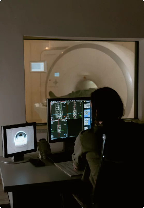
Advanced diagnostics no longer depend solely on what a doctor can see in an image. Research is now focusing on extracting all kinds of detailed and quantitative information from these images.
Traditionally, Computed Tomography (CT) has been used, but it has low sensitivity when detecting peritoneal implants smaller than one centimeter. Now, Multiparametric Magnetic Resonance Imaging (MRI) is emerging as a method capable of detecting tumor cellularity and angiogenesis.
Consequently, AI-based software is being developed to help radiologists detect focal and premalignant lesions in the abdomen (pancreas, kidney, and liver), even before the cancer has manifested. This software can identify lesions and automate long-term patient follow-up, by automatically comparing images to detect even the slightest change. This allows for a highly accurate prediction of whether a patient will respond well to preoperative chemotherapy and whether they will be a good candidate for surgery, optimizing results and avoiding unnecessary procedures.
Conclusion
It is crucial to understand that this innovation does not mean abandoning proven therapies. Cytoreductive Surgery (CRS), combined with Hyperthermic Intraperitoneal Chemotherapy (HIPEC), remains the cornerstone of patients’ treatment. But doctors are consistently working to improve patient therapies.
At PCI, we continue to train surgeons in these complex procedures, acknowledging that surgical experience is irreplaceable. The recent recognition of Dr. Domenico Sabia as mentor by the ESSO (European Society of Surgical Oncology) underscores the importance of transferring this experience to the next generation.
The combination of human experience and cutting-edge technology allows for continued improvement in the diagnosis of peritoneal cancer patients.